|
|
Normal processes
- embryonic development, fetal maturation, and perinatal changes
- organ structure and function
- chambers, valves
- cardiac cycle, mechanics, heart sounds, cardiac conduction
- Cardiac cycle:
| Sounds: |
S4 (atrial contraction) |
S1 |
|
S2 (splitting upon inspiration) |
S3 |
| Valves (which side first): |
|
AV close (L) |
AP open (R) |
AP close (L) |
AV open (R) |
| Murmurs start at: |
|
AV close = AV regurgitation, Septal |
AP open = Aortic stenosis (squirting) |
AP close = AP regurgitation |
AV open = AV stenosis (squirting) |
| Jugular vein pulse: |
a (atrial contraction) |
|
c (tricuspid bulge) |
|
v (pressure drop when AV opens/begins to fill) |
- Mechanics:
| Phase: |
4: before reaching threshold |
0: depolarize |
1: repolarize |
2: contraction |
3: repolarize |
| Pacemaker channels open: |
Na+ (If) |
Ca2+ |
|
|
K+ (DRK) |
| Ventricular channels open: |
K+ (IRK) |
Na+ |
K+ (ItoK) |
Ca2+ |
K+ (DRK) |
- Conduction:
- SA → AV (delay separates atrial and ventricular contraction) → Bundle of His → Perkinje fibers
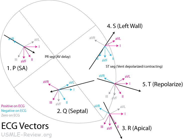
- ECG:
- PR interval = a measure of AV delay. AV block if > 0.2 sec (1 big square)
- P wave = atrial depolarization. SA dead if none seen.
- PR segment = AV delay.
- QT interval = duration of ventricular systole ~ 0.4 sec (2 big squares)
- QRS interval = ventricular depolarization. Normally < 0.1 sec (1/2 big square). Only SA, AV, and bundle of His produces normal looking QRS. Abnormal QRS = PVC, V Tach.
- ST segment = ventricles depolarized ~ 0.1 sec (1 big square)
- T wave = ventricular repolarization ~ 0.1 sec (1 big square)
- ECG vectors:
- hemodynamics, including systemic, pulmonary, coronary, and blood volume
- Venous return
- Greater return if stiffer veins (sympathetic vasoconstriction).
- Pulmonary
- Does not hold back on flow (high compliance and low resistance, lack of constrictors from myogenic, metabolic, sympathetic regulation).
- Demand driven homeostasis: divert blood where there's most oxygen (constrict hypoxic + injured areas).
- Keep pressure to minimum to prevent filtration and edema.
- Block in pulmonary backs up on right ventricle (right ventricle failure), deprives left ventricle (decrease stroke volume).
- Block in left ventricle pushes back on pulmonary (pulmonary edema).
- Heart
- Stroke volume: how much the heart squeezes out. Preload and contractility helps stroke volume. Afterload works against it.
- Preload: how much the ventricles fill (venous pressure) before contraction. Tachycardia = no time for ventricle refill = decreased preload.
- Afterload: force heart must overcome when squeezing. Increased afterload means increased diastolic pressure, stiff arteries, aortic valve stenosis.
- Contractility: how hard the heart contracts.
- Cardiac output = mean arterial pressure / resistance
- Systole: Systolic pressure increases with large stroke volume and stiff arteries.
- Diastole: Diastolic pressure increases with vasoconstriction.
- Heart rate: increased rate decreases time in diastole/ventricular filling.
- Circulation
- Resistance is greatest in arterioles and least in the vena cava.
- Resistance increases with vasoconstriction.
- Symptoms:
- Old age: increased blood pressure (both systolic and diastolic), incrased pulse pressure.
- circulation in specific vascular beds
- cell/tissue structure and function
- heart muscle, metabolism, oxygen consumption, biochemistry, and secretory function
- endothelium and secretory function, vascular smooth muscle, microcirculation, and lymph flow
(including mechanisms of atherosclerosis)
- Endothelium: release NO (vasodilation), prostacyclin (anticlot).
- Vascular smooth muscle: responds to intrinsic (myogenic, metabolic, autacoids) and extrinsic (sympathetic, parasympathetic, hormonal) regulation.
- Myogenic = flow homeostasis = constrict when pressure is high, dilate when pressure is low. Present in arteries except for pulmonary.
- Metabolic = metabolic products as vasodilators
- flow homeostasis: fast flow washes away vasodilators → vasoconstriction → decreased flow
- demand driven: high metabolism → vasodilation
- Autacoids = autacrines and paracrines.
- Vasodilators: histamine, NO, prostacyclin
- Vasoconstrictors: Thromboxane, endothelin
- neural and hormonal regulation of the heart, blood vessels, and blood volume, including responses to
change in posture, exercise, and tissue metabolism
- Homeostasis driven
- Arterial baroreflex: maintain homeostasis by modifying sympathetic and parasympathetic (vagus) system.
- baroreceptors in left and right carotid sinuses and in aortic arch. They fire in response to stretch during systole.
- baroreceptors resets: takes chronic high/low blood pressure as the norm. Consequences: works only to stabilize short-term changes in blood pressure. Eg. standing up after prolonged bed-rest causes drop in blood pressure and syncope.
- Venous baroreflex: homeostasis by modifying ADH, renin release (and to lesser extent sympathetic and vagal system).
- receptors in vein-atria junction (mostly right). They fire in response to atrial stretch during diastole.
- Renin-Angiotensin: increases blood pressure.
- Renin is released by the kidney. Catalyzes angiotensinogen → angiotensin I.
- ACE (angiotensin converting enzyme) catalyzes angiotensin I → angiotensin II.
- Cerebral ischemic reflex: raises blood pressure (sympathetic) during severe hypotension. Symptoms: unconscious patient with tachycardia maintaining low to normal bp.
- Demand driven
- Arterial chemoreflex: metabolic demand (low O2, high CO2, high H, low pH) causes sympathetic stimulation, increased respiratory rate.
- sensors in ventral medulla, carotid body, aortic body.
- Cushing reflex: try to get blood flowing in the brain when strong intracranial pressure is squeezing on the brain vessels. Symptoms: hypertension (sympathetic, both sys- and diastolic) + bradycardia (vagal drive, mechanism unknown).
- Pain, fear, anger increases sympathetic drive.
- Vasoconstrictors = NE, angiotensin, vasopressin = increase intracellular Ca2+ (Ca 2+ channels open + SR release)
- Vasodilators = Histamine, Bradykinin, Nitroglycerin, EPI, NE = decrease intracellular Ca2+ (Ca 2+ channels close + SR sequestration)
- repair, regeneration, and changes associated with stage of life
Abnormal processes
- infectious, inflammatory, and immunologic disorders
- traumatic and mechanical disorders
- neoplastic disorders
- metabolic and regulatory disorders (including dysrhythmias, systolic and diastolic dysfunction, low- and
high-output heart failure, cor pulmonale, systemic hypertension, ischemic heart disease, myocardial
infarction, systemic hypotension and shock, and dyslipidemias)
- Atrial arrhythmias
- Sinus bradycardia = everything normal, just that SA is firing slow (< 60/min, over 5 big squares per beat).
- Sinus tachycardia = everything normal, just that SA is firing fast (> 100/min, less than 3 big squares per beat).
- Premature atrial contraction (PAC) = everything normal except for an occasional premature beat.
- Atrial bigeminy = every other beat is premature.
- Paroxysmal supraventricular tachycardia (PSVT) = tachycardia without P waves.
- Atrial flutter/fibrillation = oscillations/vibrations of the baseline with normal QRS waves.
- Ventricular arrhythmias
- Premature ventricular contraction (PVC) = everything normal except an occasional ugly QRS.
- Ventricular tachycardia = tachycardia with ugly QRS.
- Ventricular fibrillation = no QRS.
- AV Blocks
- 1st degree: PR interval over 0.2 sec (1 big square)
- 2nd degree: Dropped beats: some P waves dont make it (not followed by QRS)
- Mobitz type I (Wenckebach): progressively increasing PR interval leads to dropped beat.
- Mobitz type II: dropped beats without changes in PR interval.
- 3rd degree: none of the P waves make it → AV takes over → slow, regular beats.
- vascular disorders
- systemic diseases affecting the cardiovascular system
- congenital and genetic disorders of the heart and central vessels
- idiopathic disorders
- drug-induced adverse effects on the cardiovascular system
- degenerative disorders
Principles of therapeutics
- mechanisms of action, use, and adverse effects of drugs for treatment of disorders of the
cardiovascular system
- coronary and peripheral vasodilators
- antiarrhythmic drugs
- antihypertensive drugs
- measures used to combat hypotension and shock
- drugs affecting cholesterol and lipid metabolism
- drugs affecting blood coagulation, thrombolytic agents, and antiplatelet agents
- inotropic agents and treatment of heart failure
- immunosuppressive, antimicrobial, antineoplastic, and antiparasitic drugs
- drugs to treat peripheral arterial disease
- other pharmacotherapy
- other therapeutic modalities
Gender, ethnic, and behavioral considerations affecting disease treatment and prevention, including psychosocial,
cultural, occupational, and environmental
- emotional and behavioral factors
- influence on person, family, and society
- occupational and other environmental risk factors
- gender and ethnic factors
|
|